
Epistasis: Why Two Mutations Together Don’t Just Add Up
A foundational, naive assumption in molecular biology and protein design is that the functional effects of individual mutations are strictly additive. On paper, if Mutation A reduces protein folding stability by 1.5 kcal/mol, and Mutation B reduces it by 1.0 kcal/mol, a researcher often assumes that the double mutant containing both variations should exhibit a combined destabilization of approximately 2.5 kcal/mol.
This simple, linear assumption frequently fails in practice. In complex macromolecules, combining two mutations routinely produces functional outcomes that deviate significantly from their sum. This non-additivity is not an exceptional, rare caveat: it is an inherent, structural consequence of how amino acid residues interact in continuous physical space.
This article serves as a definitive, technically precise reference guide to the epistasis definition in structural biology. We examine its mathematical definition relative to null models, analyze its physical and mechanistic origins, and outline how non-additivity is quantified and mapped across modern protein engineering pipelines.
Understanding Epistasis Classifications
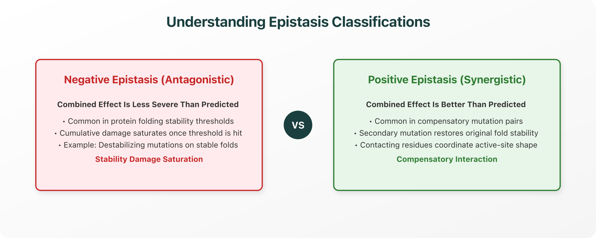
The non-linear interactions between mutations fall into distinct statistical and physical categories, depending on whether their combined effect is more or less severe than predicted:
1. Defining Epistasis Mathematically
In genetics and biophysics, epistasis is formally defined as the statistical deviation between the observed phenotypic effect of a double mutant and the effect predicted by combining the individual effects of the single mutants under a specific null expectation model.
The Null Models:
- The Additive Null Model: Typically applied when measuring thermodynamic properties (such as folding free energy change, ΔΔG):
ε = ΔΔG_AB − (ΔΔG_A + ΔΔG_B)
Where ε represents the epistatic coefficient. If ε = 0, the mutations are perfectly additive. Any deviation (ε ≠ 0) indicates epistasis.
- The Multiplicative Null Model: Standardly applied in evolutionary biology and fitness studies (where W represents organismal or protein fitness):
ε = W_AB − (W_A × W_B)
This mathematical framing highlights a critical conceptual point: epistasis is not an absolute, intrinsic property of a sequence; it is defined relative to a specific null expectation model.
2. The Mechanistic Origins of Epistasis
Non-additivity in proteins is driven by specific, well-defined biophysical and functional communication networks:
- Biophysical Stability Margins: Most natural proteins are only marginally stable, typically possessing a folding stability buffer (ΔG_f) of 5 to 15 kcal/mol. If Mutation A and Mutation B both destabilize the fold, their cumulative damage can push the protein past its threshold, driving denaturation. Once the folding threshold is crossed, further destabilizing mutations have no additional measurable effect because the protein is already unfolded, resulting in negative, saturating epistasis.
- Functional Pathway Epistasis: Within a signaling cascade or biochemical pathway, enzymes act in series. If Mutation A completely inactivates the primary upstream enzyme, the functional impact of a secondary downstream Mutation B is completely masked. The effect of the second mutation depends entirely on whether the pathway is already disrupted by the first.
- Protein-Structural Context and Contacting Residues: When two mutated residues physically contact each other in 3D space, their interaction is highly direct. For example, if Mutation A destroys a critical salt bridge (e.g., mutating an aspartate to alanine), it causes severe destabilization. However, if a neighboring Mutation B simultaneously restores that charge-charge interaction (e.g., mutating a lysine to glutamate), it functions as a compensatory mutation, restoring the local structural environment and rescuing folding stability.
3. Positive vs. Negative Epistasis
Understanding the direction of non-additivity is essential for predicting variant profiles:
- Negative (Antagonistic) Epistasis: Occurs when the combined effect of the two mutations is more severe than predicted (in fitness contexts) or when cumulative destabilizing damage saturates because the protein has already unfolded.
- Positive (Synergistic) Epistasis: Occurs when the double mutant performs significantly better than expected under the null model. This is commonly observed in compensatory mutation pairs, where a secondary variation physically rescues the structural defect introduced by the first.
4. How Epistasis Is Quantified
Modern biochemistry utilizes two primary experimental workflows to map non-additive landscapes:
Double Mutant Cycle Analysis
The classic standard for measuring the coupling energy (ΔΔG_int) between two specific residues:
- The Workflow: Researchers express and purify four variants: the wild type (WT), the single mutant A, the single mutant B, and the double mutant AB.
- The Calculation: By measuring the thermodynamic stability of all four variants, they calculate the exact thermodynamic coupling energy:
ΔΔG_int = (ΔG_AB − ΔG_B) − (ΔG_A − ΔG_WT)
This value defines the direct physical and cooperative interaction energy between the two target loci.
Deep Mutational Scanning (DMS)
While double mutant cycles are limited to checking single pairs, deep mutational scanning DMS allows for the systematic, high-throughput mapping of epistatic interactions across thousands of mutation pairs simultaneously.
- The Workflow: DMS utilizes massive synthetic variant libraries to express thousands of double-mutant combinations in a single pool, applying selective pressure (such as binding selection or cell survival), and utilizing high-throughput sequencing to quantify the abundance of every variant before and after selection.
- The Output: This generates comprehensive, high-resolution epistatic landscape maps, showing exactly where mutations cooperate or conflict across the entire protein structure.
5. Practical Implications for Protein Design and Genomics
Acknowledging and modeling epistasis is a non-negotiable requirement across active discovery workflows:
- Protein Engineering Constraints: When optimizing an enzyme for thermal stability or binding affinity, combining five individually beneficial mutations rarely produces a hyper-stable variant. Due to local structural conflicts and stability saturation, the mutations often exhibit negative epistasis. Rational design requires testing combinations and utilizing joint structural models, rather than assuming independent additivity.
- Predicting Variant Effects in Genomics: In clinical genomics and target curation, interpreting compound variants (multiple mutations on the identical gene, often termed haplotypes) is a major challenge. The clinical impact of a disease-associated mutation can be completely masked or severely amplified by the presence of a second, background variation, meaning clinical interpretation must evaluate the complete genetic background, not just individual variants in isolation.
Closing: A Structural Feature of Biology
For researchers and genome engineers, the core take-away is clear:
- Additivity is a mathematical simplification that requires rigorous justification in any macromolecular study.
- Epistasis is the structural, biophysical standard of how mutations actually combine.
By transitioning from linear assumptions to systematic, coordinate-level epistatic mapping (utilizing double mutant cycles and DMS datasets), structural biology teams can predict variant effects with absolute thermodynamic accuracy, paving the way for rational, highly robust protein design.
References and Authoritative Specifications
For structural biologists seeking to inspect the thermodynamic models and high-throughput datasets discussed, the following publications serve as primary references:
- The Double Mutant Cycle Method: Carter, P. J. et al. (1984). "The use of double mutant cycles in protein engineering." Cell, 38(3), 835-840. doi:10.1016/0092-8674(84)90278-8
- Deep Mutational Scanning of Epistasis: Fowler, D. M., & Fields, S. (2014). "Deep mutational scanning: a new generation of quantitative genetics." Nature Methods, 11(8), 801-807. doi:10.1038/nmeth.3027
- Biophysical Principles of Protein Epistasis: Starr, T. N., & Thornton, J. W. (2016). "Epistasis in protein evolution." Protein Science, 25(7), 1204-1218. doi:10.1002/pro.2897
- Protein Stability Folding and Saturation: Tokuriki, N., & Tawfik, D. S. (2009). "Stability effects of mutations and protein evolvability." Current Opinion in Structural Biology, 19(5), 596-604. doi:10.1016/j.sbi.2009.08.003
Explore Purna's Molecular Intelligence Platform
AI-powered workspace for biology teams to accelerate drug discovery from target identification to lead optimization.
Try Purna AI →